
Cervical cancer begins in the cervix, which is the lower part of the uterus, commonly known as the womb. The womb, or uterus, is a vital organ in the female reproductive system. It is positioned in the pelvis, between the bladder and the rectum. Specifically, the cervix acts as the narrow passage connecting the uterus to the vagina, often referred to as the birth canal.
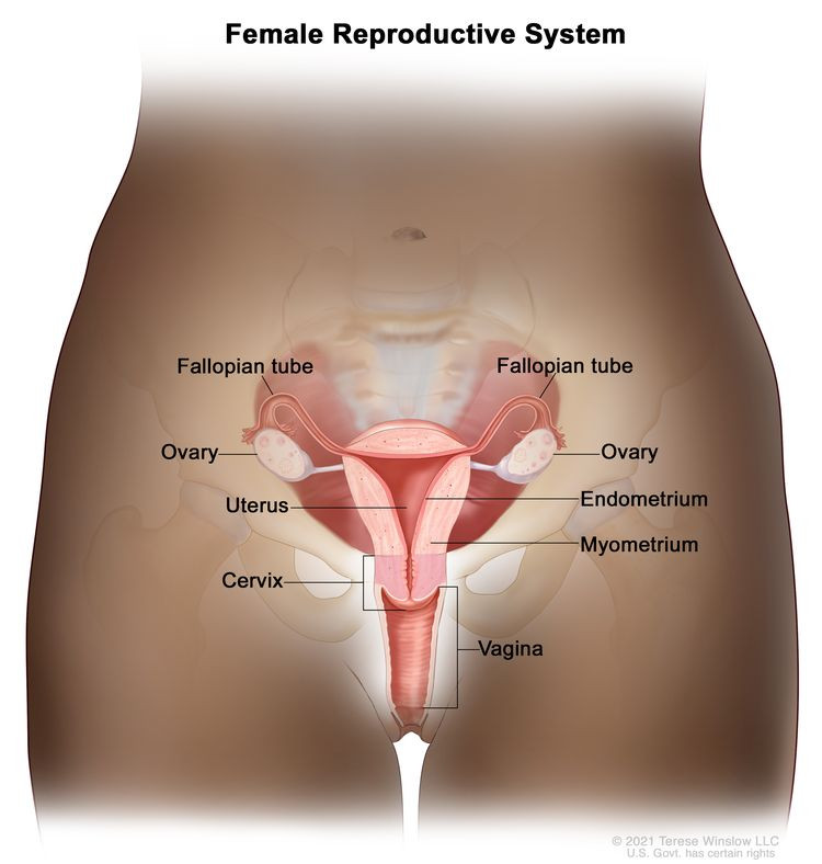 Anatomy of the female reproductive system illustrating the position of the womb
Understanding the anatomy is crucial in grasping how cervical cancer develops. The cervix itself has distinct parts. The squamocolumnar junction, also known as the transformation zone, is where two types of cells meet – the endocervix and ectocervix. Most cervical cancers originate in this zone. Before cancer develops, cells in the cervix may undergo changes called dysplasia, leading to abnormal cells in the cervical tissue.
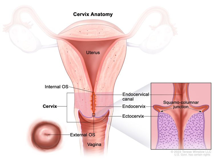 Detailed anatomy of the cervix highlighting the transformation zone
If these abnormal cells are not addressed, they can evolve into cancer cells. These cancerous cells can then grow and spread deeper into the cervix and potentially to surrounding areas. Therefore, knowing “Where Is The Womb” and understanding the structure of the cervix is the first step in comprehending cervical cancer and its development within the female reproductive system.