Your heart, a vital organ responsible for pumping life-sustaining blood throughout your body, is centrally located within your chest. Understanding exactly where your heart is situated and its basic anatomy is crucial for appreciating its function and maintaining cardiovascular health. This article will explore the precise location of the heart, its protective structures, and key anatomical features.
Pinpointing the Heart’s Location in Your Chest
The heart resides in the chest cavity, more specifically within a region known as the mediastinum. This area is situated between your lungs, placing the heart in a relatively central and protected location. While many people believe the heart is on the left side of the chest, it’s actually positioned more towards the middle, slightly tilted to the left.
To be precise, your heart is located behind and slightly to the left of your breastbone, also known as the sternum. You can feel your sternum as the bony plate in the center of your chest. This sternum provides a degree of protection to the heart, along with the rib cage.
 Anatomical illustration showing where the heart is situated in the chest, positioned behind the sternum and between the lungs.
Protective Layers: The Pericardium
The heart isn’t simply floating freely in your chest; it’s enclosed within a protective sac called the pericardium. This pericardium is a double-layered membrane that surrounds the heart, acting like a protective barrier.
The outer layer of the pericardium is tough and fibrous, attaching to major blood vessels connected to the heart and anchoring it to surrounding structures like the spinal column and diaphragm. The inner layer closely adheres to the heart muscle itself. Between these two layers is a fluid-filled space. This pericardial fluid acts as a lubricant, allowing the heart to move smoothly and contract efficiently as it beats, minimizing friction against surrounding tissues.
Size and Weight: The Heart’s Physical Dimensions
Despite its powerful function, the heart is a relatively compact organ. An adult human heart is approximately the size of a clenched fist. In terms of weight, it typically ranges from 7 to 15 ounces (200 to 425 grams).
Consider the incredible workload of this fist-sized organ. Over an average lifetime, a heart may beat more than 3.5 billion times. On a daily basis, the average heart beats around 100,000 times, diligently pumping approximately 2,000 gallons (7,571 liters) of blood throughout the body.
Inside the Heart: Chambers and Septum
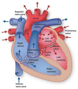
The heart is divided into four chambers, which work in coordination to circulate blood. The upper chambers are called the atria (singular: atrium), specifically the left atrium and the right atrium. These atria receive blood returning to the heart.
The lower chambers are the ventricles, namely the left ventricle and the right ventricle. These ventricles are responsible for pumping blood out of the heart. A muscular wall, known as the septum, separates the left and right atria and the left and right ventricles, ensuring that oxygen-rich and oxygen-poor blood remain separate.
The left ventricle is the largest and most muscular chamber. While its walls are only about half an inch thick, they generate enough force to propel oxygenated blood through the aortic valve and into the aorta, the body’s largest artery, for distribution to the entire body.
The Heart Valves: Regulating Blood Flow
To ensure unidirectional blood flow through the heart, four valves are present. These valves act like one-way doors, preventing backflow and maintaining efficient circulation:
- Tricuspid valve: Controls blood flow between the right atrium and the right ventricle.
- Pulmonary valve: Regulates blood flow from the right ventricle into the pulmonary arteries, which carry deoxygenated blood to the lungs for oxygenation.
- Mitral valve: Allows oxygen-rich blood from the lungs to flow from the left atrium into the left ventricle.
- Aortic valve: Opens to permit oxygen-rich blood to pass from the left ventricle into the aorta, the main artery supplying blood to the body.
The Heart’s Electrical System: The Conduction System
The rhythmic beating of the heart is controlled by an intrinsic electrical system known as the conduction system. Electrical impulses originating within the heart muscle (myocardium) trigger heart contractions.
This electrical signal generation begins in the sinoatrial (SA) node, located in the right atrium. The SA node is often referred to as the heart’s natural pacemaker. The electrical impulse from the SA node spreads through the atria, causing them to contract, and then travels to the ventricles, causing them to contract and pump blood. While the SA node establishes a baseline heart rate, factors like physical activity, stress, and hormones can influence and adjust heart rate as needed.
The Heart’s Role in the Circulatory System
The heart and the circulatory system together form the cardiovascular system. The heart functions as a powerful pump, propelling blood to all organs, tissues, and cells throughout the body. This blood delivery is essential for providing oxygen and vital nutrients to every cell and for removing carbon dioxide and waste products generated by cellular activity.
Blood travels away from the heart through a network of arteries, smaller arterioles, and tiny capillaries, where oxygen and nutrient exchange occurs. Deoxygenated blood returns to the heart through venules and veins, completing the circulatory loop. Remarkably, if all the blood vessels in the body were laid end-to-end, they would stretch for approximately 60,000 miles (over 96,500 kilometers), enough to circle the Earth more than twice, highlighting the extensive network the heart tirelessly serves.
In conclusion, your heart is situated centrally in your chest, protected by the sternum and rib cage, and enclosed within the pericardium. Understanding its location and basic anatomical features provides a foundation for appreciating the heart’s complex and vital role in maintaining overall health and well-being.