Your heart, the vital engine of life, is centrally located in your chest, playing a crucial role in your overall well-being. But Where Is Your Heart Situated exactly? It’s a common question, and understanding the precise location and anatomical details of your heart is key to appreciating its function. This article will delve into the heart’s position within your body, its size, structure, and the intricate systems it powers.
Located in the middle of your chest, your heart resides between your lungs, nestled behind and slightly to the left of your breastbone, also known as the sternum. Imagine a central compartment in your chest, and that’s where you’ll find this remarkable organ. To be more precise, it’s often described as being in the mediastinum, the space in the chest between the pleural cavities that contain the lungs.
To protect this precious organ, nature has provided a double-layered sac called the pericardium. Think of the pericardium as a protective bag that snugly envelops your heart. The outer layer of this sac extends to cover the roots of the heart’s major blood vessels and is anchored by ligaments to your spinal column, diaphragm, and other surrounding structures. This anchoring provides stability and keeps the heart in its correct position.
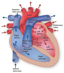 Diagram of the heart's position in the chest
The inner layer of the pericardium is closely attached to the heart muscle itself. Between these two layers is a lubricating fluid. This fluid is essential as it allows the heart to move smoothly within the pericardial sac as it beats, minimizing friction and wear and tear.
While the question “where is your heart situated” addresses location, understanding its size gives further context. The heart, though incredibly powerful, is surprisingly compact. It typically weighs between 7 to 15 ounces (200 to 425 grams), which is only a little larger than the size of your clenched fist. Despite its modest size, the heart is a tireless worker. Over an average lifetime, it may beat more than 3.5 billion times. On a daily basis, your heart beats around 100,000 times, diligently pumping approximately 2,000 gallons (7,571 liters) of blood throughout your body.
Delving deeper into heart anatomy, you’ll discover it’s composed of four chambers. These chambers are divided into upper and lower sections. The two upper chambers are known as the left and right atria. These atria are primarily receiving chambers, collecting blood returning to the heart. Below them are the two lower chambers, the left and right ventricles. The ventricles are the powerful pumping chambers that propel blood out to the lungs and the rest of the body.
A muscular wall, called the septum, acts as a divider, separating the left and right atria, as well as the left and right ventricles. This separation is crucial to prevent the mixing of oxygen-rich and oxygen-poor blood, ensuring efficient oxygen delivery to your body.
Notably, the left ventricle stands out as the largest and strongest of the four chambers. Although its walls are only about half an inch thick, the left ventricle generates enough force to push blood through the aortic valve and into the aorta, the body’s largest artery, initiating systemic circulation.
The Heart Valves: Gatekeepers of Blood Flow
Within these chambers are four vital valves that meticulously regulate the flow of blood through your heart. These heart valves act like one-way doors, ensuring blood moves in the correct direction and preventing backflow.
- The Tricuspid Valve: This valve controls blood flow between the right atrium and the right ventricle. It opens to allow deoxygenated blood to flow from the right atrium into the right ventricle and closes to prevent backflow during ventricular contraction.
- The Pulmonary Valve: Positioned between the right ventricle and the pulmonary arteries, the pulmonary valve governs blood flow from the right ventricle into the pulmonary arteries. These arteries carry blood to your lungs where it picks up oxygen.
- The Mitral Valve: Also known as the bicuspid valve, the mitral valve manages the flow of oxygen-rich blood from your lungs, allowing it to pass from the left atrium into the left ventricle.
- The Aortic Valve: This valve is the gateway for oxygen-rich blood to exit the left ventricle and enter the aorta. The aorta is the body’s largest artery, distributing oxygenated blood to the entire systemic circulation.
The Conduction System: The Heart’s Electrical Grid
What orchestrates the rhythmic beating of your heart? It’s an intricate electrical system known as the conduction system. Electrical impulses originating from your heart muscle, the myocardium, trigger heart contractions. This electrical signal journey begins in the sinoatrial (SA) node, often referred to as the heart’s “natural pacemaker,” located in the top of the right atrium.
The SA node spontaneously generates electrical impulses at a regular rhythm. This electrical impulse then spreads throughout the muscle fibers of the atria and ventricles, causing them to contract in a coordinated manner. While the SA node sets a baseline heart rate, your actual heart rate is remarkably adaptable, changing in response to physical demands, stress levels, and hormonal influences.
The Circulatory System: The Heart’s Network
The heart is not an isolated organ; it’s the central component of your cardiovascular system, which also includes the extensive circulatory system. Your heart functions as a powerful pump, propelling blood to every organ, tissue, and cell in your body. This blood is not just a fluid; it’s a delivery system, carrying oxygen and vital nutrients to each cell while simultaneously removing carbon dioxide and waste products generated by cellular activity.
Blood embarks on a journey from your heart to the body via a complex network of arteries, arterioles, and capillaries. Arteries are the major highways, arterioles are smaller roads, and capillaries are the tiny, branching pathways that reach individual cells. After delivering its cargo and picking up waste, blood returns to your heart through venules and veins, completing the cycle.
The extent of this circulatory network is astonishing. If you were to lay out all the blood vessels in your body end-to-end, they would stretch for approximately 60,000 miles (over 96,500 kilometers). That’s a distance long enough to circle the Earth more than twice! This vast network underscores the heart’s critical role in sustaining life by ensuring every part of your body receives the oxygen and nutrients it needs to function.
Understanding where your heart is situated and its intricate anatomy provides a profound appreciation for this incredible organ and its tireless work in keeping you alive and well.