Understanding where your heart is located and how it’s structured is fundamental to appreciating its vital role in your overall health. This powerful organ, the core of your circulatory system, works tirelessly to sustain life. So, Where Is The Heart Located within your body? Let’s delve into the anatomy of this incredible muscle and explore its position and key components.
Your heart is positioned centrally within your chest, nestled between your lungs in a region known as the mediastinum. To be more precise, it sits behind and slightly to the left of your breastbone, or sternum. This placement offers a degree of protection while still allowing the heart to function effectively. Enveloping the heart is a protective sac-like structure called the pericardium, a double-layered membrane. The outer layer of the pericardium anchors to the roots of the heart’s major blood vessels and is connected via ligaments to your spinal column, diaphragm, and other parts of your anatomy, providing stability and preventing excessive movement.
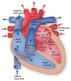 Diagram of the heart's location in the chest cavity
In terms of size, your heart is remarkably compact, typically only a little larger than your clenched fist and weighing between 7 to 15 ounces (approximately 200 to 425 grams). Despite its size, the heart’s workload is immense. Over an average lifetime, it may beat more than 3.5 billion times. On a daily basis, this translates to around 100,000 beats, pumping an astonishing volume of approximately 2,000 gallons (7,571 liters) of blood throughout your body.
Delving deeper into the heart’s structure, you’ll find it’s composed of four chambers, crucial for efficient blood circulation. The upper chambers are termed the left and right atria, while the lower, more muscular chambers are the left and right ventricles. A robust muscular wall, known as the septum, acts as a divider, separating the left and right atria, and similarly, the left and right ventricles. Notably, the left ventricle stands out as the heart’s largest and most powerful chamber. Despite its walls being only about half an inch thick, the left ventricle generates enough force to propel blood through the aortic valve and into the systemic circulation, delivering oxygenated blood to the entire body.
To ensure unidirectional blood flow through these chambers, the heart relies on four essential valves. These valves act like gates, opening and closing in coordination with the heart’s contractions. The tricuspid valve manages blood flow between the right atrium and the right ventricle. The pulmonary valve governs the movement of blood from the right ventricle into the pulmonary arteries, which are responsible for carrying deoxygenated blood to the lungs for oxygenation. The mitral valve facilitates the passage of oxygen-rich blood from the lungs, directing it from the left atrium into the left ventricle. Finally, the aortic valve controls the outflow of oxygen-rich blood from the left ventricle into the aorta, the body’s largest artery, initiating systemic circulation.
The heart’s rhythmic contractions are driven by an intrinsic electrical system known as the conduction system. Electrical impulses originating within the heart muscle itself, the myocardium, trigger these contractions. This electrical signaling begins in the sinoatrial (SA) node, often referred to as the heart’s “natural pacemaker,” situated in the top region of the right atrium. From the SA node, electrical impulses propagate through the muscle fibers of both the atria and ventricles, prompting them to contract in a coordinated manner. While the SA node establishes a baseline heart rate, various factors such as physical activity, stress levels, and hormonal influences can modulate heart rate to meet the body’s changing needs.
The heart, in conjunction with the circulatory system, constitutes the cardiovascular system, a vital network for life support. The heart functions as a powerful pump, propelling blood to every organ, tissue, and cell in your body. This blood supply is crucial for delivering oxygen and essential nutrients while simultaneously removing carbon dioxide and metabolic waste products. Blood travels away from the heart through a complex network of arteries, arterioles, and capillaries, reaching even the most remote corners of the body. The deoxygenated blood then returns to the heart via venules and veins, completing the circulatory loop. If you were to lay out all the blood vessels in your body end-to-end, they would stretch for an astounding 60,000 miles (over 96,500 kilometers), a distance sufficient to circle the Earth more than twice!
In conclusion, understanding where is the heart located and its basic anatomy provides crucial insight into its function as the central organ of the circulatory system. Its position in the chest, its chambers, valves, and electrical system all work in harmony to ensure the continuous circulation of blood, delivering life-sustaining oxygen and nutrients throughout your body. Appreciating this intricate design underscores the importance of maintaining a healthy heart for overall well-being.